Klaus Ersfeld
Laboratory of Molecular Parasitology
Department of Genetics
University of Bayreuth
95440 Bayreuth
Germany
Klaus Ersfeld
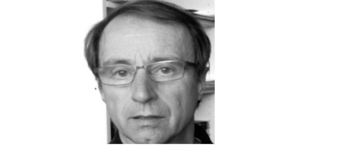
...studied biology and geography at the University of Bonn (1981-87) and did his PhD at the Max-Planck-Institute for Biophysical Chemistry in Göttingen cloning (big deal in those days) the first enzyme involved in tubulin post-translational modifications, the tubulin-tyrosine ligase. Inspired by a parasitology course as an undergraduate student, he decided to move into the area of molecular parasitology and worked as a postdoc at the Liverpool School of Tropical Medicine on a project developing a diagnostic assay for tapeworm infections based on recombinant proteins. This also involved a short stint working in Kenya. In 1994 he joined the lab of Keith Gull at the University of Manchester and, initially as a postdoc then as an independent group leader, worked on aspects of genome organisation and cytoskeletal biology of Trypanosoma brucei. In 2003, he was appointed Senior Lecturer at the University of Hull in northern England and continued work on the cytoskeleton of trypanosomes, focusing on the role of kinesin motorproteins. Since 2011, he is Professor of Molecular Parasitology at the University of Bayreuth.
Research synopsis
The cytoskeleton of trypanosomes is defined by a corset of subpellicular microtubules. We investigate how the dynamics of these microtubule arrays are regulated and contribute to morphological adaptations during the life cycle of the parasite. A particular focus is on the role of microtubule post-translational modifications, such as polyglutamylation. This is a joint project with the Department of Experimental Physics (Prof. Matthias Weiss), funded by the DFG priority program "Physics of Parasites". We use a variety of molecular biology techniques, such as mutagenesis, epitope tagging, gene deletions and RNA interference. Equally important are, however, cell biology techniques to probe the 3-dimensional structure of the cells. Techniques such as immunofluorescence microscopy, advanced digital image analysis (cell tracking, motility analysis), and electron microscopy are routinely employed.
This research is Project 5 of the SPP 2332 PoP.


